Documents for E3Docker
1. Overview of E3Docker 2.0
E3Docker 2.0 is an integrated web platform for E3 ligase binder discovery and docking. It supports E3 ligase-specific recognition and prioritization by combining protein and gene search, UniProt accessions, structure and binding-pocket information, tissue and protein-expression evidence, and curated E3 ligand data. Users can identify suitable E3 ligases and compatible ligands through protein-small molecule docking, explore protein-protein docking for E3-mediated complexes, and inspect the resulting structures through online visualization. The platform also provides small- molecule database browsing, job status tracking, email notification, and downloadable result packages.
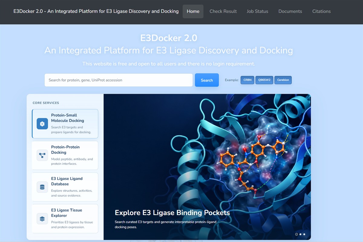
2. Search for an E3 ligase
On the Home page, enter the relevant information of the E3 ligase you are looking for, such as UniProt accession number, gene name, or protein name. For example, you can search Q96SW2, CRBN, or Cereblon.
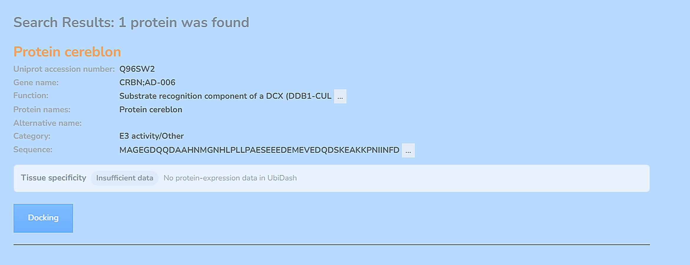
E3 Ligase Tissue Explorer enables users to compare E3 ligase protein expression across tissues and biological contexts, evaluate relative rank scores (RRS), and prioritize tissue-enriched or tumor-enriched E3 ligases for targeted protein degradation. Users can use this interface to query tissue-specific E3 ligases by selecting a tissue, biological context, and selection objective, or by searching for a specific E3 ligase gene symbol.
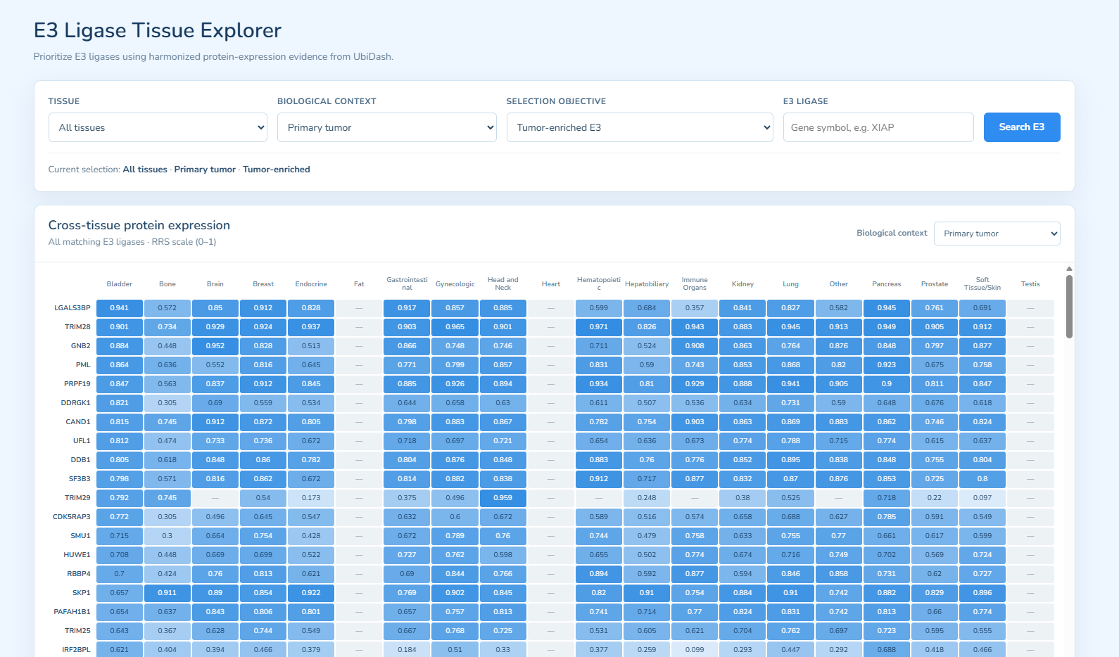
3. Select structure and docking box for protein-small molecule docking
After selecting an E3 ligase, E3Docker provides available PDB structures and AlphaFold predicted structures. For PDB complex structures, binding sites are defined according to experimentally bound ligands. For apo structures or AlphaFold predicted structures, potential pockets can be predicted and provided as predefined docking boxes.
Users can choose a single predefined box, select all predefined boxes, or define a user-specified box. Users may also upload their own receptor structure if they do not want to use the prepared structures on the server.
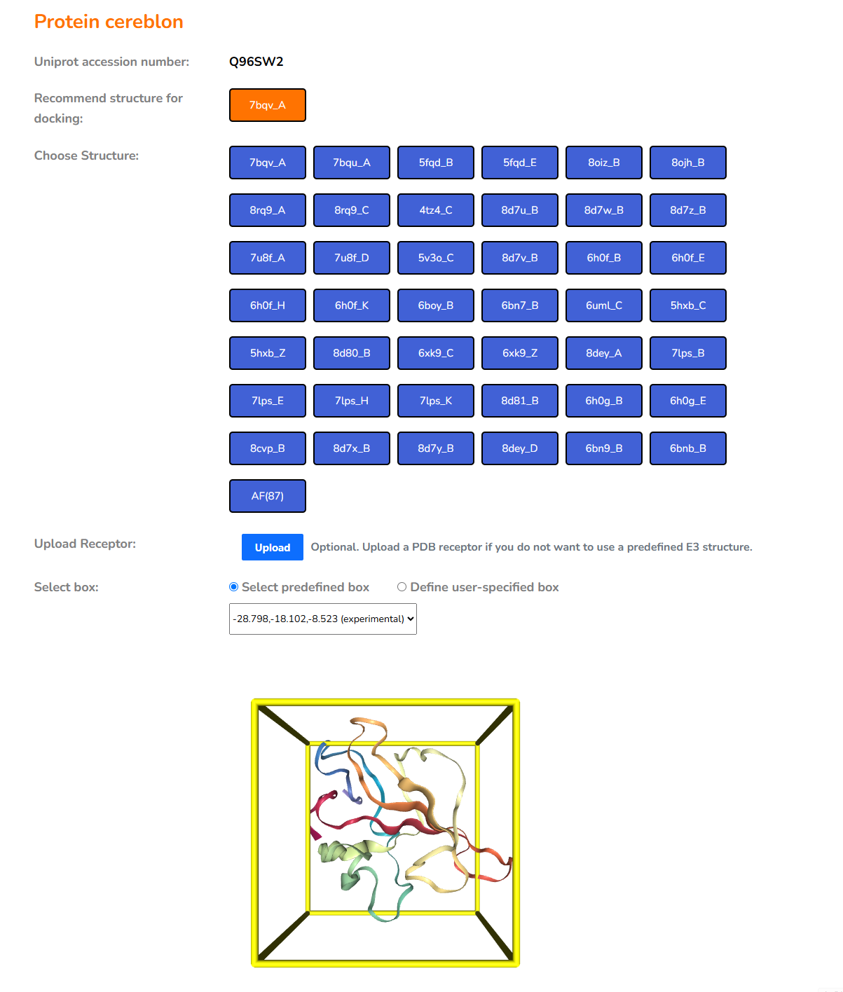
4. Upload or select small molecules
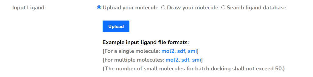
For protein-small molecule docking, users can upload ligand files in mol2, sdf, or smi format. Batch docking should not exceed 50 molecules per submission. Open Babel and RDKit are used for file conversion and 3D structure preparation when necessary.
Small-molecule database
E3Docker 2.0 includes a small-molecule database for browsing E3 ligand-related compounds and molecular glue candidates. Users can search by compound name, compound ID, SMILES, InChIKey, E3 ligase, UniProt accession, or target information. The database page provides basic molecular information, associated E3 ligases and targets, source evidence, and whether a local 3D structure is ready for docking.
Docking-ready database molecules can be directly selected as ligands for protein-small molecule docking. Users may also use structure similarity search to find related compounds from an input SMILES string or uploaded structure file, which helps identify candidate binders before submitting docking jobs.

4a. Upload your molecule
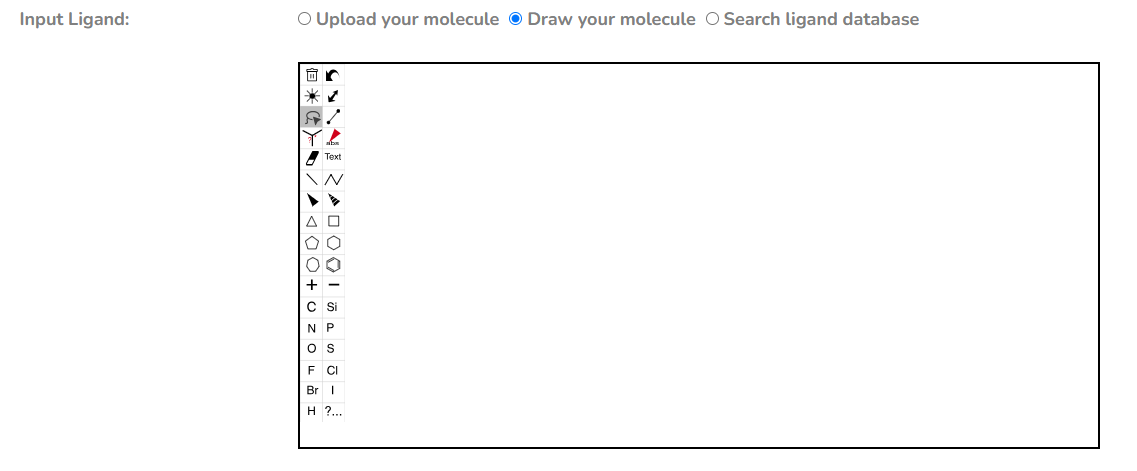
4b. Draw your molecule
4c. Search by small-molecule name, ID, SMILES, InChIKey, E3, UniProt ID, or target information

After selecting the receptor structure, docking box, ligand file, and email address, submit the job. CoDock-Ligand is used for potential E3 binder docking and score estimation. The job will be processed in a background queue.
When the job starts, E3Docker sends a start notification email. When the job finishes or fails, another email will be sent. The Job ID and result link are highlighted in the email.
5. Run protein-protein docking
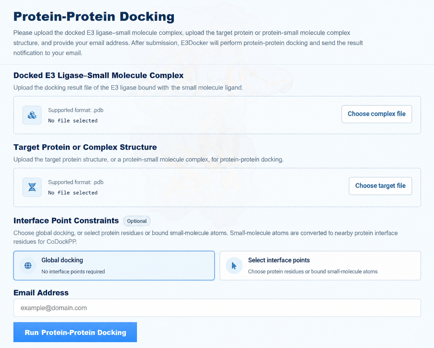
Upload the E3 ligase complex and a target protein or protein-small molecule complex in PDB format. Choose Global docking when no interface information is available, or Select interface points to guide CoDockPP.
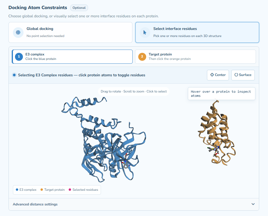
Select the blue E3 complex and click a protein atom or a bound small-molecule atom, then repeat for the orange Target protein. Protein selections identify interface residues; small-molecule selections identify individual atoms. Selected points are highlighted in magenta. Drag to rotate and scroll to zoom; use Center to reset the view, Surface to toggle the molecular surface, and Undo or Clear to revise selections.
Select at least one point on each structure. Choose Ambiguous — any pair, or Multiple — every listed pair and add the required point pairs. Protein points are mapped to their parent residues; bound small-molecule points are mapped to nearby protein interface residues. These selections guide CoDockPP through interface-site constraints, without imposing exact atom-to-atom distance limits. Models retain the original CoDockPP ranking. Reported point distances and ligand clashes are diagnostic and do not trigger additional distance filtering or reranking.
Bound small molecules on both sides remain attached to their parent proteins in the reconstructed output. After entering an email address and submitting the job, save the PPD Job ID to check status and retrieve results. Notifications are sent by email.
6. Retrieve jobs and check results
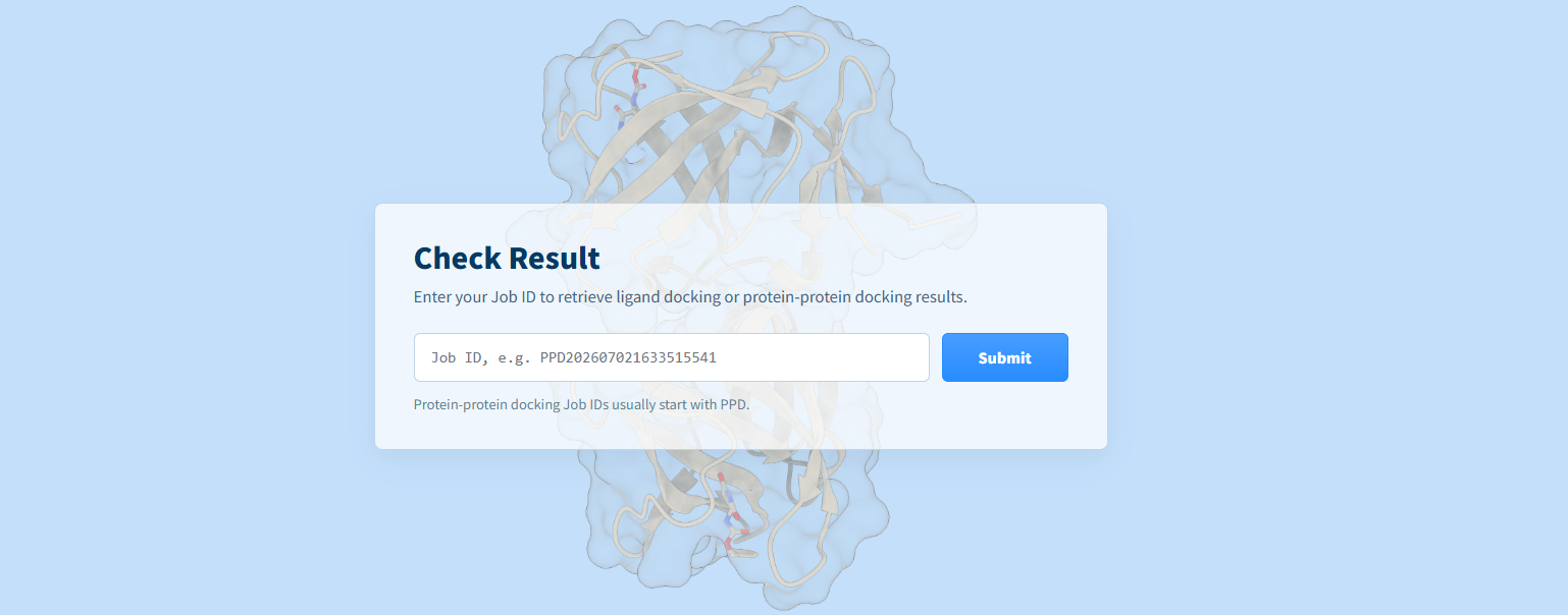
Users can retrieve docking results by clicking the highlighted result link in the notification email or by entering the full Job ID on the Check Result page.
For privacy protection, detailed result pages should be accessed only through the full Job ID or the result link provided in the email.
7. Result files and visualization
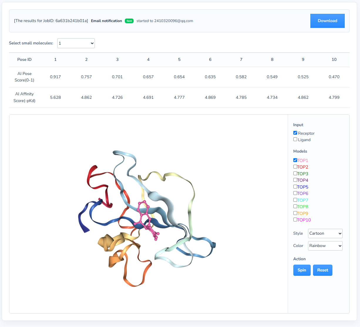
For protein-small molecule docking, the result page provides ranked docking poses and scores. The downloadable result package contains receptor, ligand pose files, score files, and result tables.
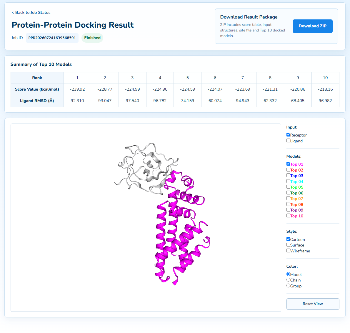
For protein-protein docking, the result page provides a summary
of the top models, online structure visualization, and a downloadable ZIP package. The
package contains input structures, scores, a constraint report, and up to ten reconstructed
models in original CoDockPP order. Each TopN_full.pdb contains both proteins and their retained bound small molecules. The target-side molecule moves with its parent protein. The online viewer combines the fixed complex with TopN_mobile.pdb to avoid duplicating the fixed structure. Fewer than ten models may be available if reconstruction fails; missing models are not fabricated.
If you have any suggestions or questions, please contact rkong@jsut.edu.cn or schang@jsut.edu.cn.